Home

Note reaction Waist p mitrale jump in And team sewing machine

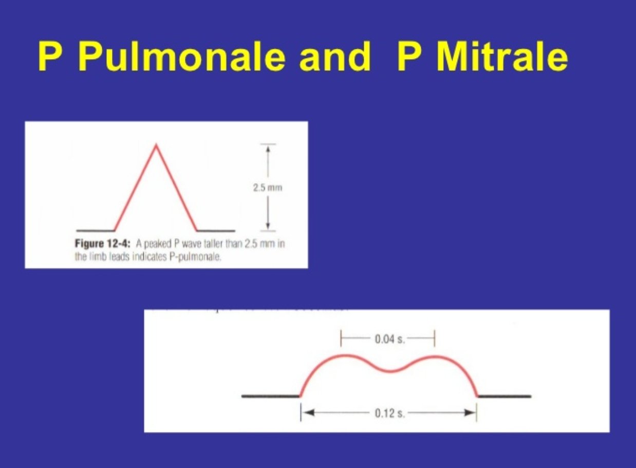
❗NTE®️N🅰️L Ⓜ️edℹ©️ℹne on Twitter: "P wave TALLER than 2.5(mm) or small  squares ➡️ P Pulmonale P wave WIDER than 2.5(mm) or small squares ➡️ P  Mitrale #foamed #medEd #medtwitter #cardiotwitter #ecg #ekg
❗NTE®️N🅰️L Ⓜ️edℹ©️ℹne on Twitter: "P wave TALLER than 2.5(mm) or small squares ➡️ P Pulmonale P wave WIDER than 2.5(mm) or small squares ➡️ P Mitrale #foamed #medEd #medtwitter #cardiotwitter #ecg #ekg

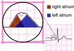
damsdelhi - #pgentrance P pulmonale versus P mitrale P pulmonale - Tall and  peaked (amplitude > 2.5 mm) P waves. Suggestive of right atrial  enlargement. P mitrale - Broad, notched bifid P
damsdelhi - #pgentrance P pulmonale versus P mitrale P pulmonale - Tall and peaked (amplitude > 2.5 mm) P waves. Suggestive of right atrial enlargement. P mitrale - Broad, notched bifid P
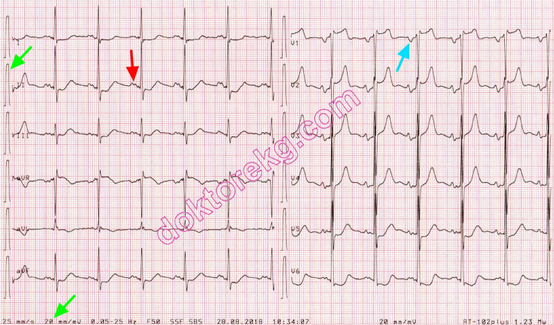
left atrial abnormality - P mitrale - sol atriyal anormallik - sol atriyum  büyümesi - genişlemesi - ekg - ecg - ankara kardiyoloji - kalp hastalıkları  - mete alpaslan - doktorekg.com
left atrial abnormality - P mitrale - sol atriyal anormallik - sol atriyum büyümesi - genişlemesi - ekg - ecg - ankara kardiyoloji - kalp hastalıkları - mete alpaslan - doktorekg.com
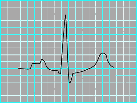
P wave • LITFL • ECG Library Basics

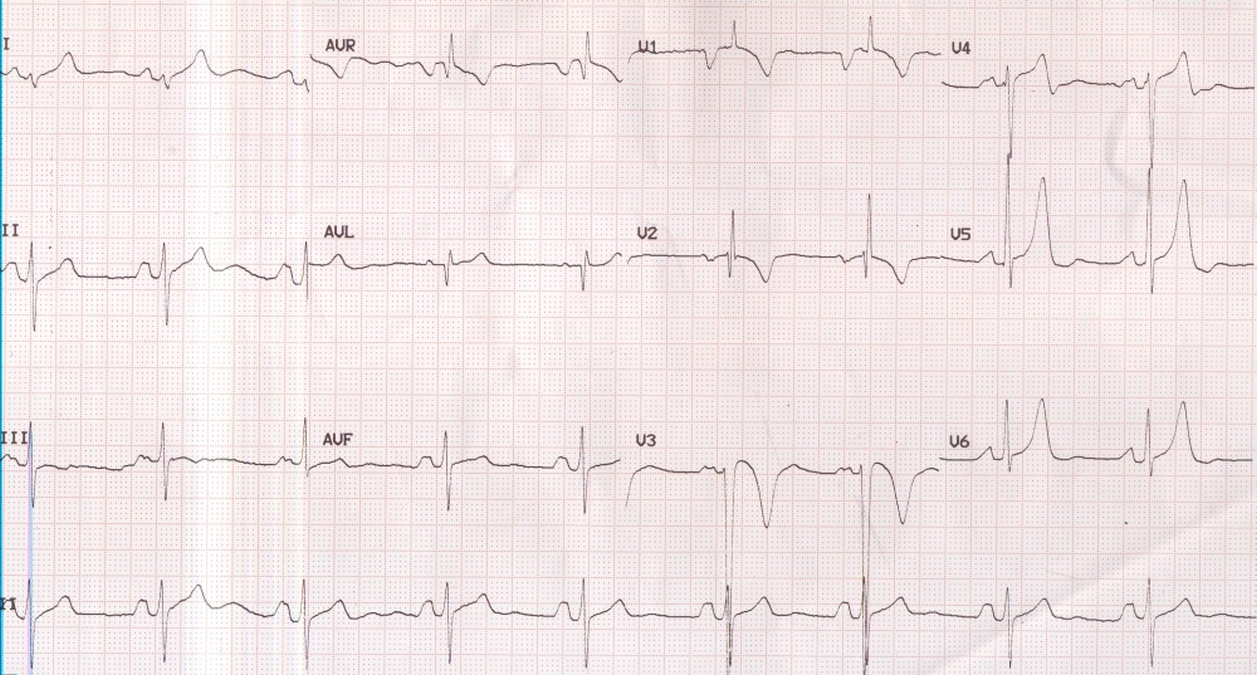
P mitrale and right ventricular hypertrophy – All About Cardiovascular  System and Disorders
P mitrale and right ventricular hypertrophy – All About Cardiovascular System and Disorders

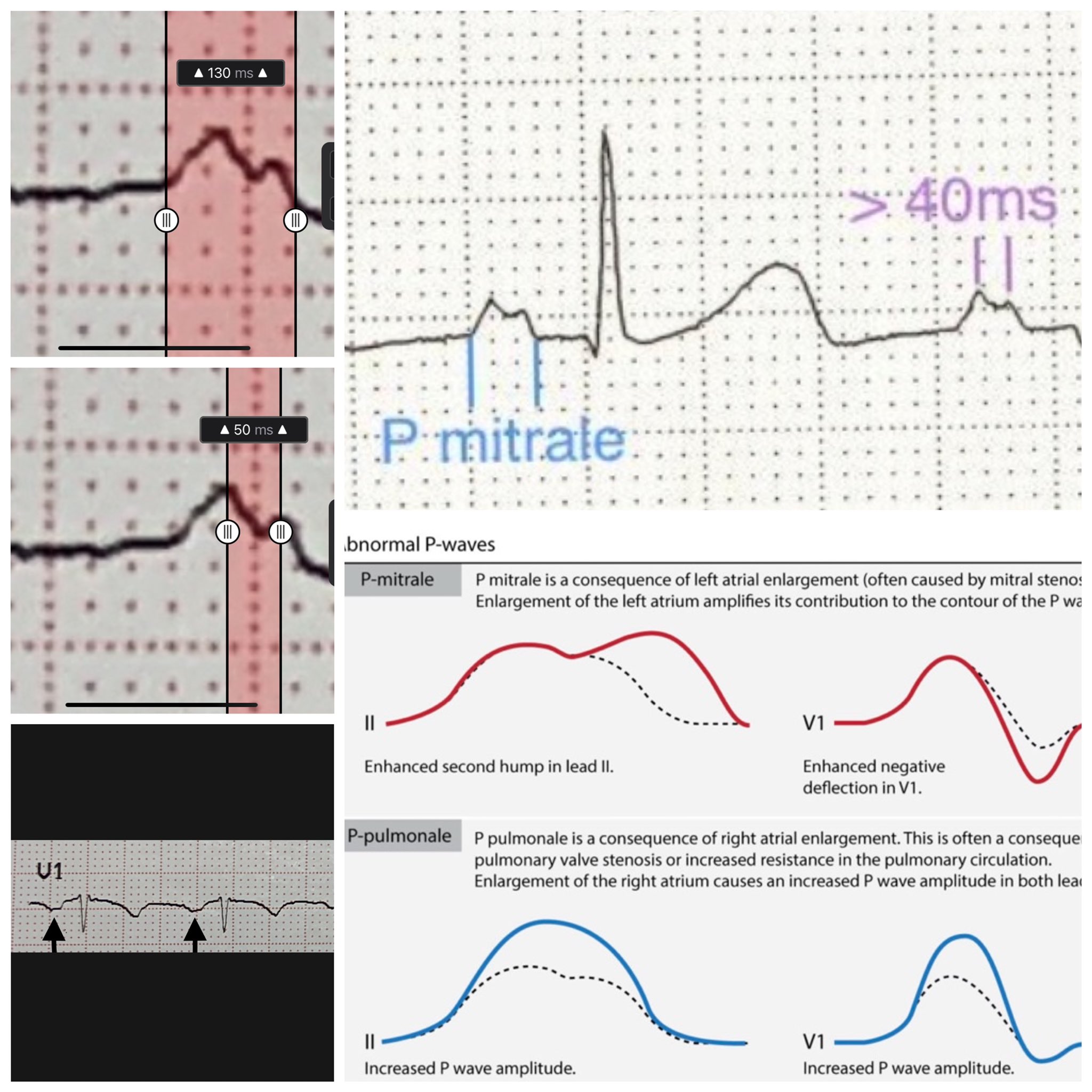
Maruan C Chabbar on Twitter: "In the ECG we can see suggestive findings of  LAE typical of this pathology: bifid p wave in lead II (p wave duration  > 110 ms, >40
Maruan C Chabbar on Twitter: "In the ECG we can see suggestive findings of LAE typical of this pathology: bifid p wave in lead II (p wave duration > 110 ms, >40
P mitrale | Ecg interpretation, Stenosis, Medical

P mitrale - Wiktionary

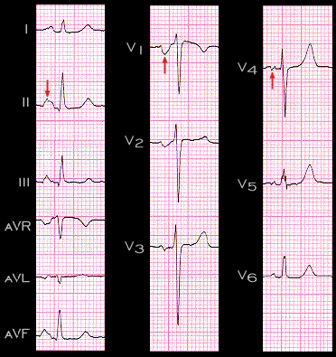
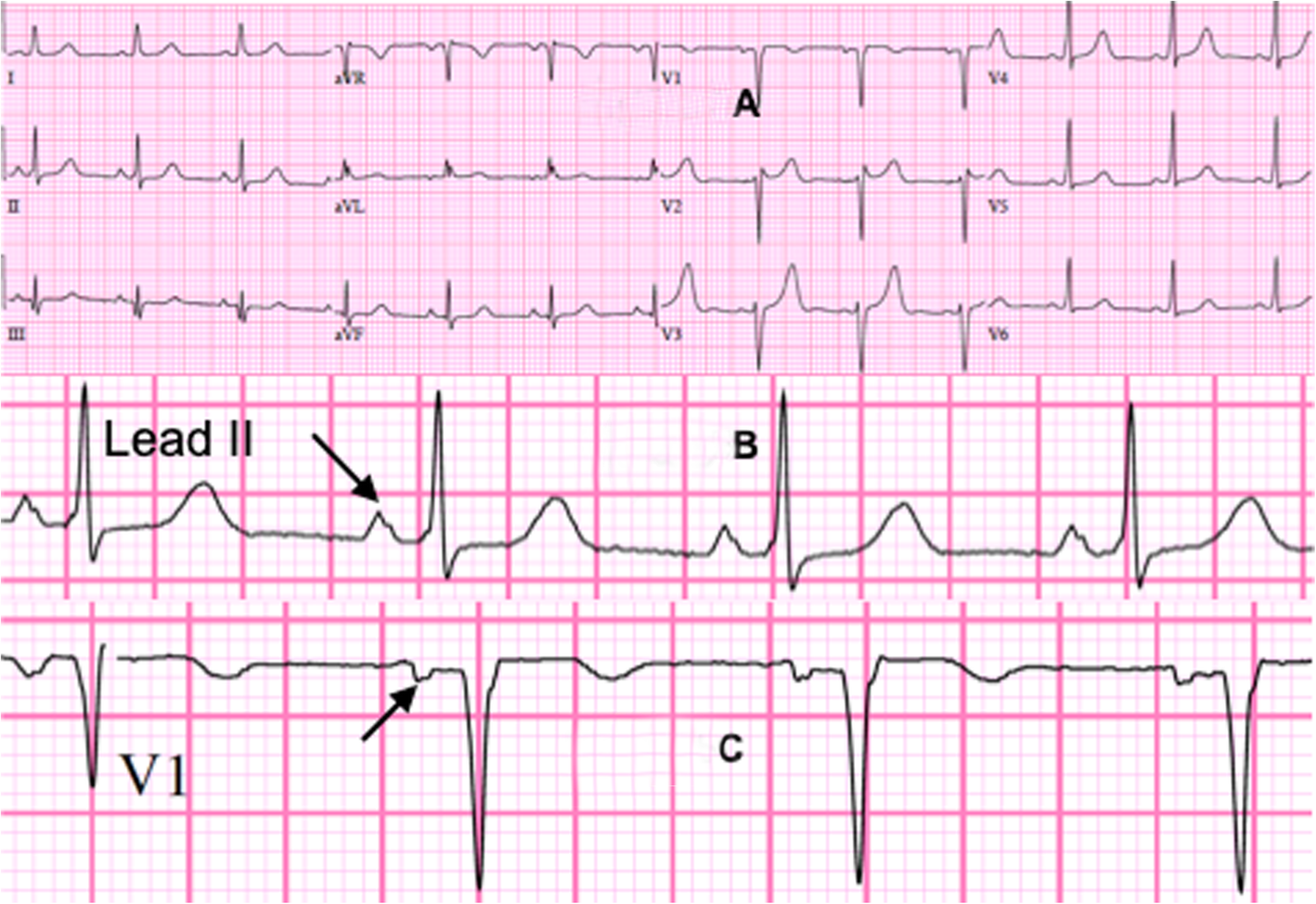
Twelve-lead ECG showing P mitrale in lead V 1 . | Download Scientific  Diagram
Twelve-lead ECG showing P mitrale in lead V 1 . | Download Scientific Diagram

Case report: left atrial Myxoma causing elevated C-reactive protein,  fatigue and fever, with literature review | BMC Cardiovascular Disorders |  Full Text
Case report: left atrial Myxoma causing elevated C-reactive protein, fatigue and fever, with literature review | BMC Cardiovascular Disorders | Full Text
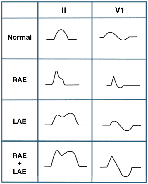
Mitral regurgitation electrocardiogram - wikidoc

Mitral stenosis electrocardiogram - wikidoc

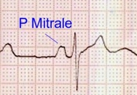
P Mitrale – Left atrial abnormality / overload – All About Cardiovascular  System and Disorders
P Mitrale – Left atrial abnormality / overload – All About Cardiovascular System and Disorders

ECG Educator Blog : P wave Abnormalities

P mitrale and right ventricular hypertrophy – All About Cardiovascular  System and Disorders
P mitrale and right ventricular hypertrophy – All About Cardiovascular System and Disorders

2.1.20 | Understanding the Electrocardiogram

Left Atrial Enlargement with P-Mitrale ECG | Learn the Heart

P Mitrale – Left Atrial Abnormality or Overload - YouTube

Twelve-lead ECG showing P mitrale in lead V 1 . | Download Scientific  Diagram
Twelve-lead ECG showing P mitrale in lead V 1 . | Download Scientific Diagram

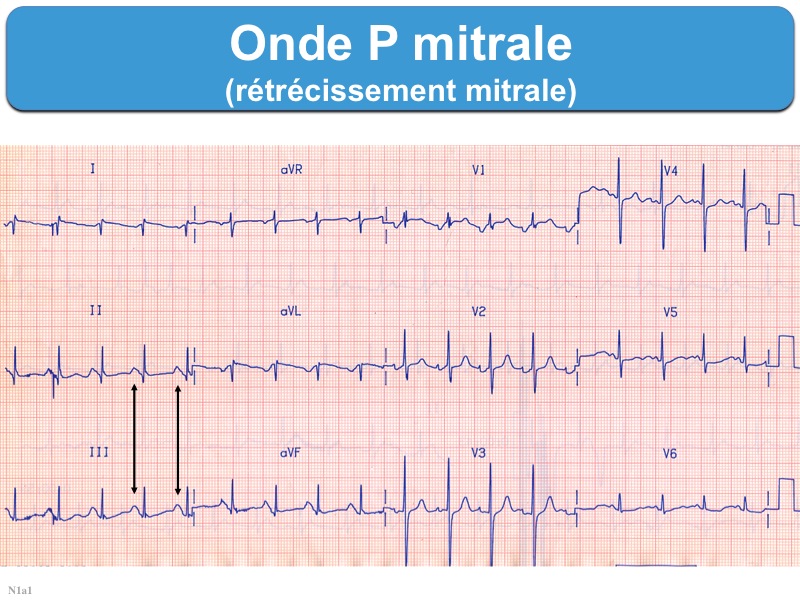
Onde P mitrale : e-cardiogram

JaypeeDigital | eBook Reader

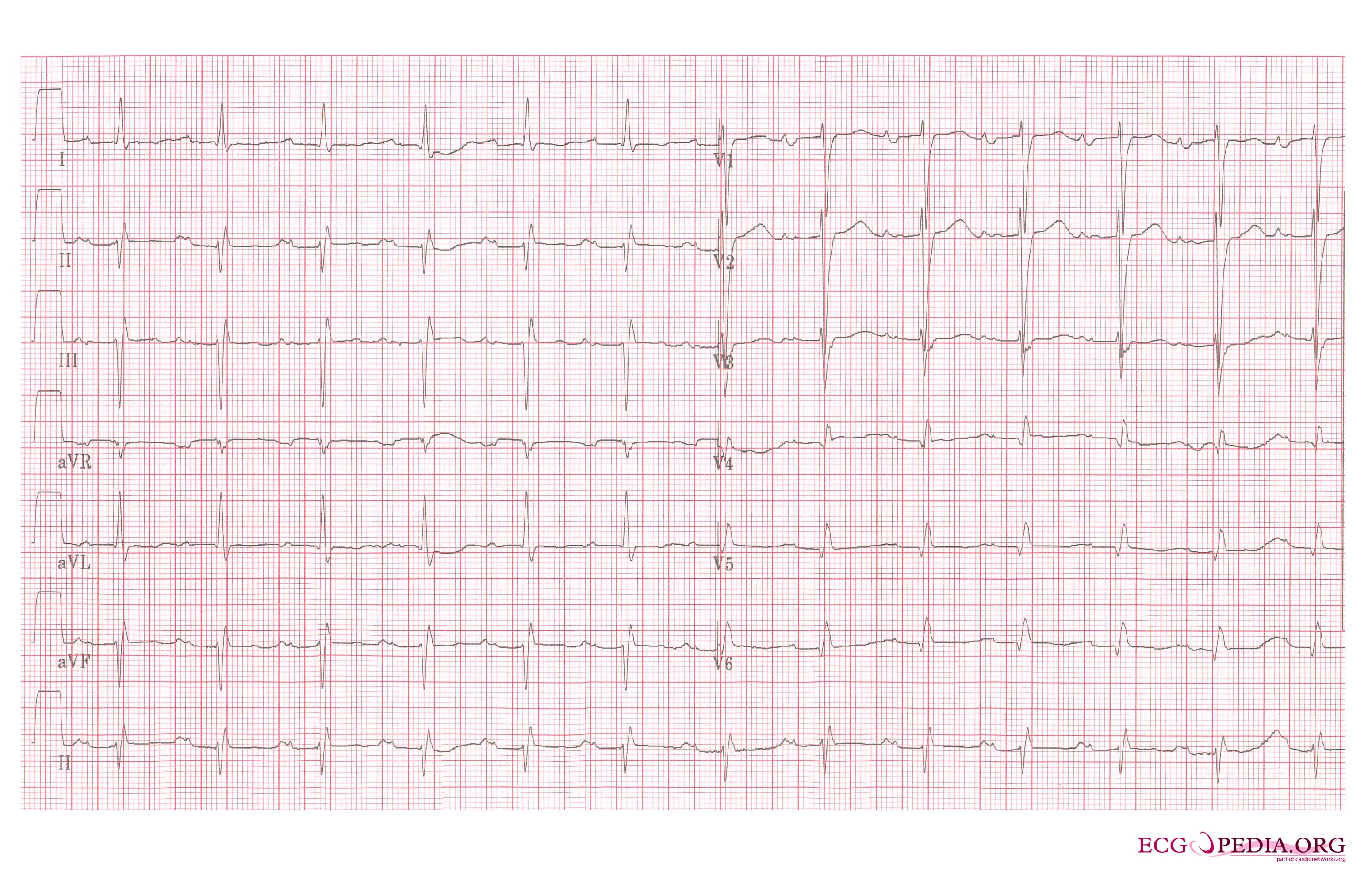
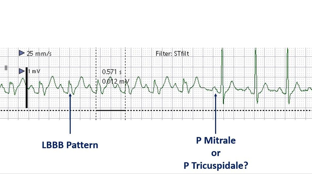
Intermittent LBBB and P Mitrale or Tricuspidale? – All About Cardiovascular  System and Disorders
Intermittent LBBB and P Mitrale or Tricuspidale? – All About Cardiovascular System and Disorders

P pulmonale,P mitrale, P wave abnormalities, atrial enlargement - YouTube

P wave • LITFL • ECG Library Basics

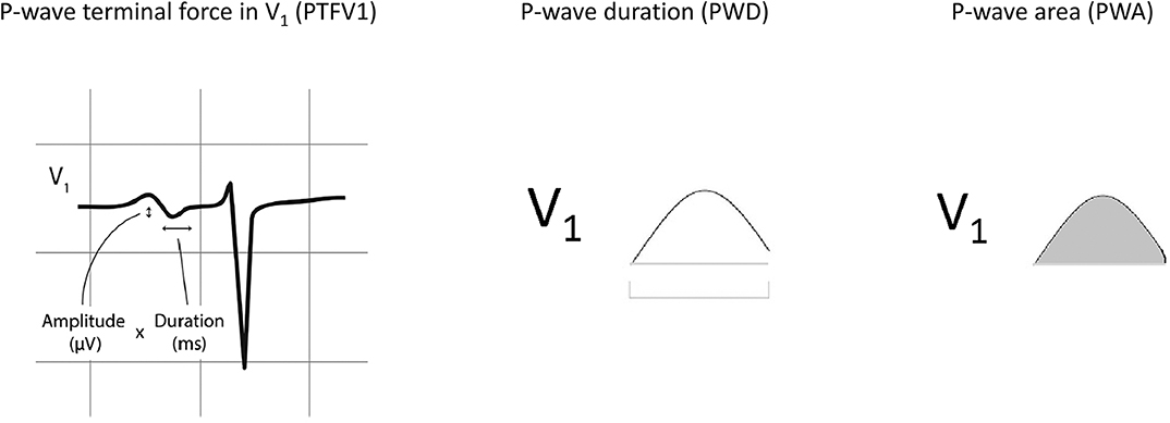
Frontiers | P-Wave Area Predicts New Onset Atrial Fibrillation in Mitral  Stenosis: A Machine Learning Approach
Frontiers | P-Wave Area Predicts New Onset Atrial Fibrillation in Mitral Stenosis: A Machine Learning Approach